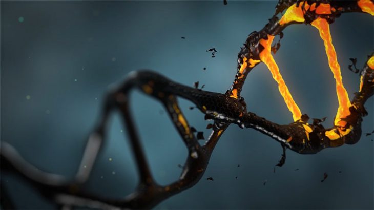
Our DNA is a crucial part of creating every cell in our bodies. It is the instruction manual our cells read to know exactly what to do and how to act. But how do our bodies proofread these instructions and know how to fix them if something goes wrong? Biochemists at the University of Utah are seeking to answer this question, and have made interesting new discoveries into the chemistry that leads to effective DNA repair.
We have 4 bases to all our DNA. They are molecules named adenine (A), guanine (G),cytosine (C), and thymine (T). Guanine is the most susceptible to a type of damage called oxidative stress, caused by rogue oxygen compounds called reactive oxygen species (ROS). ROS are a normal part of every cell’s environment. However, at high levels, certain ROS can wreak havoc on normal cell function.
In the case of guanine, ROS react by attaching an oxygen molecule to the eighth carbon in the guanine molecule, creating a new molecule called 8-oxo-guanine, or OG for short. This might not seem like much, as regular guanine and OG look really similar. However, this difference could make it difficult, or even impossible, to use this part of the DNA the way it was meant to be.
Lucky for us, our bodies have certain proteins whose job is to correct oxidative damage to our DNA. One protein in particular, known as MutY, helps initiate the process of turning 8-oxo-guanine back to regular guanine. It does this by taking away adenine (A) molecules that have accidentally bonded to OG. When guanine gets changed to OG, it creates a space perfectly shaped for adenine to fit in like a puzzle piece and attach itself to the OG molecule. These adenine molecules make it more difficult to access and repair the oxo-guanine to get back to the normal DNA sequence.
But how does MutY know to take away ONLY the adenine molecules that are bonded to OG? Regular guanine and OG look similar, and the adenine is no different from other adenine molecules. In the process of answering this question, scientists at the University of Utah discovered interesting chemical interactions between OG and MutY that could add to our understanding of how our bodies’ help repair potentially harmful DNA mutations.
In order to understand what makes OG so special to the MutY protein, the Horvath lab at the University of Utah first used different methods to learn about the structure of the MutY protein, DNA containing OG, and DNA containing regular guanine.
To start the process, they grew crystal formations of each of these molecules. The crystals were observed using a method called X-ray crystallography. It involves hitting crystals with x-rays to watch how these rays bend when they hit the molecules. X-rays bounce off of atoms in different but predictable ways called diffraction patterns, and can tell us a lot about the chemical makeup of the molecule we want to understand.
Once the scientists had a rough idea of what each molecule looked like, they used software to get an even clearer image of the two DNA molecules and the MutY protein. These computer programs are able to help interpret the x-ray diffraction patterns and turn them into detailed 3D models of the molecules.
They saw that a building block of MutY, called serine, interacted with OG in a special way. There were two extra hydrogen bond interactions between MutY and OG near this serine molecule. Hydrogen bonds are fairly strong connections between the positive end of one molecule and the negative end of another, like tiny magnets. Once these two places connect, the MutY protein is able to perform its action — removing that adenine.
To confirm their suspicion, they altered the MutY protein by substituting this serine with other molecules. They then compared how often mutations happened in E. coli cells with normal MutY and the MutY without this particular serine. They found that altering this serine and neighboring building blocks caused up to eleven times more mutations, indicating that these interactions are crucial to MutY’s ability to do its job in fixing the mutations.
While understanding just a couple more interactions between MutY and OG might seem like a small step, this discovery could be incredibly important to developing treatments to diseases associated with oxidative stress to DNA. Ovarian, colon, and pancreatic cancer have all been associated with this type of damage, along with neurodegenerative diseases like Alzheimer’s and Parkinson’s. Understanding how our body fights these diseases could help further treatments that mimic our own bodies’ first line of defense.


